
Step 01
MR image acquisition
The patient’s MR data is transferred into the planning workflow.
OrthoAlign AI
Personalized knee implant planning
OrthoAlign AI transforms MR images into patient-specific knee arthroplasty plans through anatomical segmentation and 3D bone reconstruction. Surgeons can review and adjust planning parameters in the same workspace.
A health technology R&D project supported by TÜBİTAK 1812 BiGG
OrthoAlign AI·3D planning
R&D workflow
Image processing, 3D modelling and planning come together in one digital workflow.
Step 01
The patient’s MR data is transferred into the planning workflow.
Step 02
AI separates the femur and tibia structures within the MR image.
Step 03
Segmentation data is transformed into a patient-specific three-dimensional knee model.
Step 04
Anatomical measurements, resection levels and component parameters are planned together.
Step 05
The surgeon reviews the plan, adjusts its parameters and approves it.
Planning workspace
Anatomical measurements, component sizes and positions, resection levels and alignment options are displayed together in the same workspace.
Measure key anatomical references on the femur and tibia.
Review component size, position and anatomical fit together.
Compare resection levels and related measurement parameters.
Evaluate rotation, slope and mechanical-alignment parameters.
Planning outputs
The 3D anatomical model and planning parameters create shared planning data for patient-specific cutting blocks, navigation and robotic-surgery infrastructure.
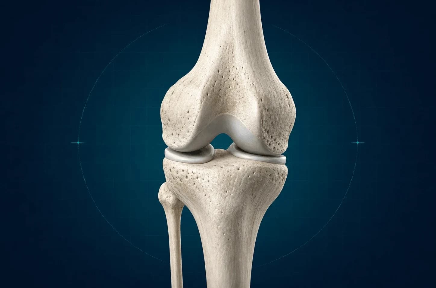
Planning output
Displays patient anatomy, measurements and planning references on an interactive 3D model.
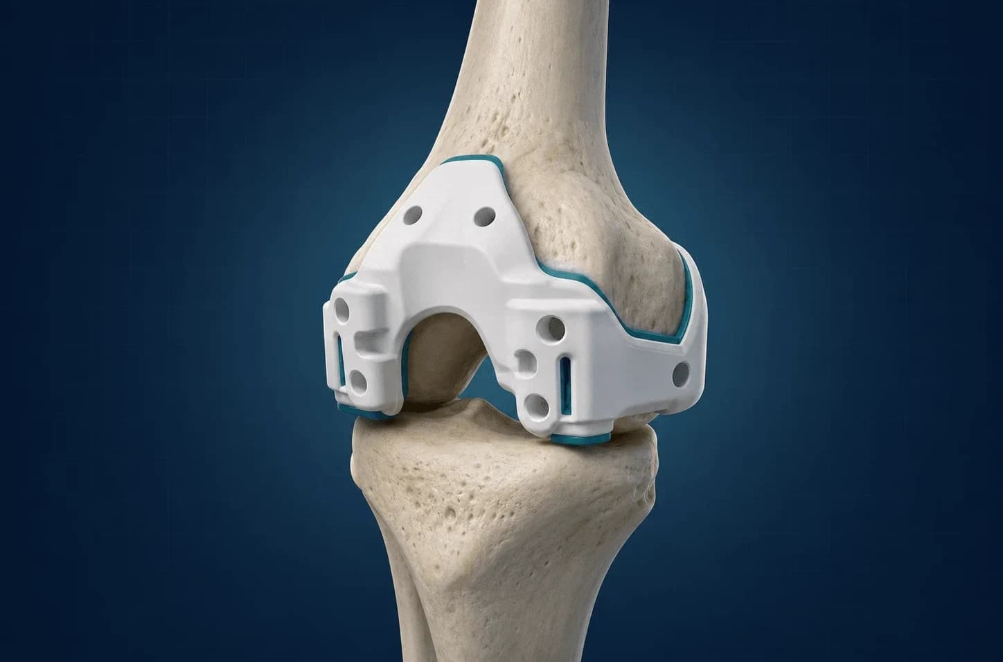
Development area
Creates the geometric data needed to design patient-specific femoral and tibial cutting guides.
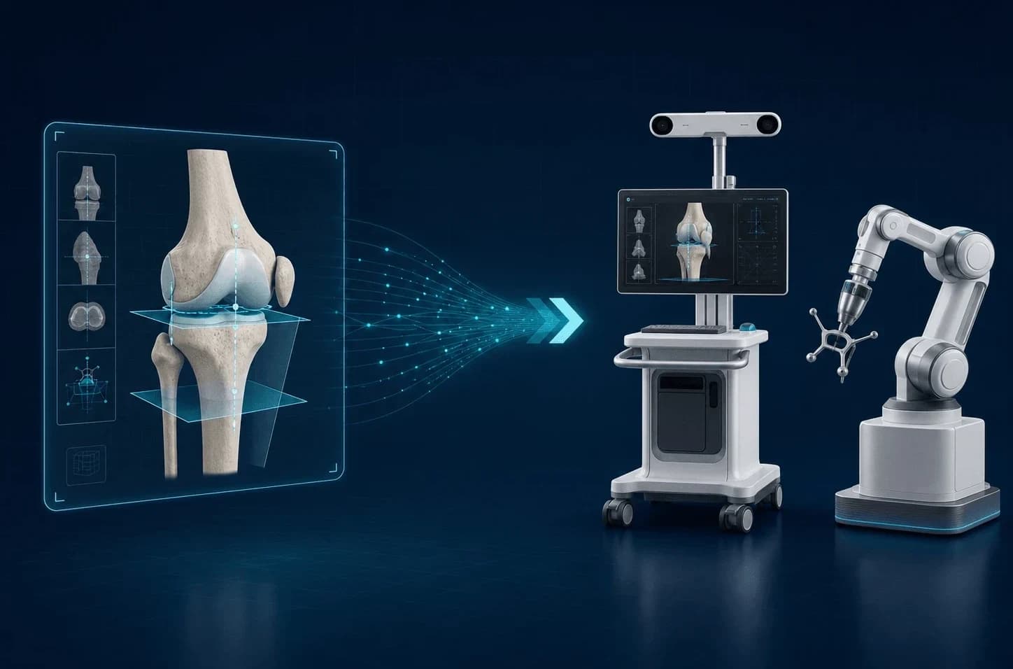
Integration research
Provides the data infrastructure for transferring planning parameters to navigation and robotic-surgery systems.
AI + surgeon expertise
OrthoAlign AI makes anatomical measurements and planning options visible. The surgeon compares and adjusts parameters to create the patient-specific plan.
3D planning experience
Femoral and tibial anatomy is displayed together with landmarks, measurement planes, alignment and component options.
View the spatial relationship between the femur and tibia from different angles.
Evaluate anatomical landmarks, axes and measurement planes in the same view.
Interactively adjust alignment, component position and resection parameters.
Scientific and technical foundation
OrthoAlign AI brings surgical-planning experience, MR image-processing research and 3D-modelling technologies together in the same product-development process.
Anatomical measurements and planning parameters are structured around real surgical workflows.
Automatic segmentation of the femur and tibia creates patient-specific 3D anatomical models.
Anatomical data becomes a measurable, comparable and interactive surgical-planning workspace.