
01
Ultrasound image
Newborn hip ultrasound image is transferred to the system for evaluation.

GrafMesh AI
Graf hip ultrasonography · Newborn hip screening
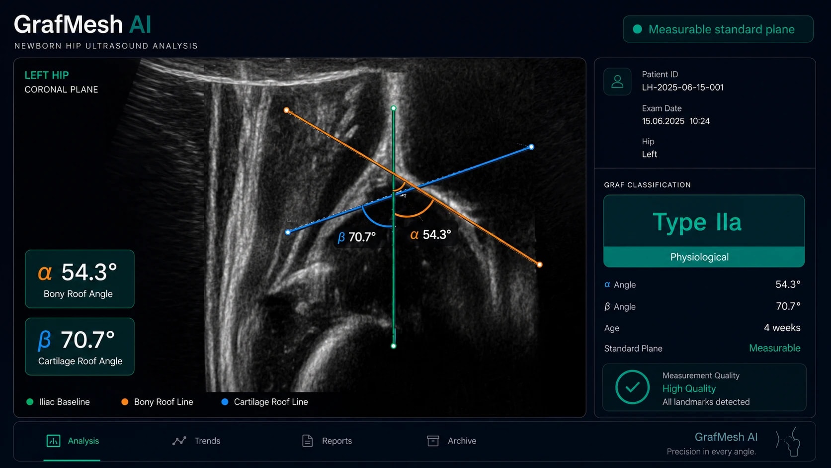
Capturing the standard plane correctly is the most critical and error-prone step in hip ultrasonography. GrafMesh AI identifies anatomical landmarks, automatically calculates α and β angles, and presents measurements alongside Graf classification.
GrafMesh AI·Graf ultrasound analysis
Product supported by the TÜBİTAK 2232-A International Leading Researchers Programme
End-to-end process
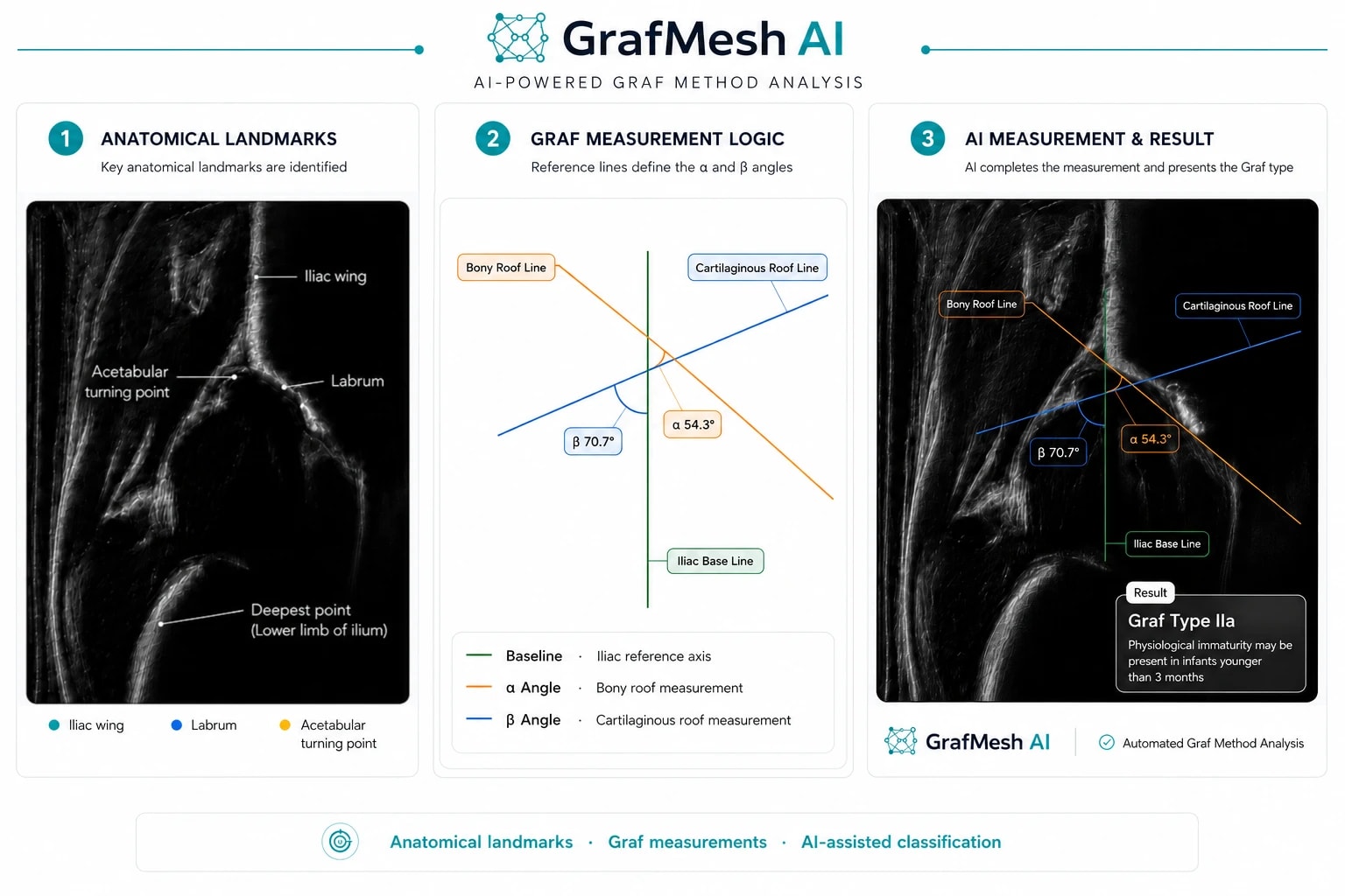
Graf evaluation begins with determining whether the image is in a standard plane suitable for measurement. Anatomical landmarks are then identified, α and β angles are calculated, and the resulting measurements are mapped to Graf classification. GrafMesh AI executes all these steps automatically.
Newborn hip ultrasound image is transferred to the system for evaluation.
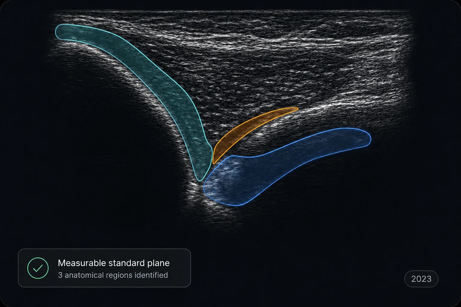
The straight appearance of the iliac wing, lower limb of the ilium, and labrum are evaluated to determine the image's suitability for Graf measurement.
The iliac wing, labrum, and necessary acetabular references are automatically identified by artificial intelligence.
α and β angles are automatically calculated based on the lines established from anatomical landmarks.
The Graf type is determined by evaluating the measurements and relevant clinical parameters, and a summary of results is presented.
Standardization in Graf assessment
Selecting a measurable standard plane, accurately identifying anatomical landmarks, and reducing inter-observer variability are essential for the consistency of Graf measurements. GrafMesh AI supports this process with shared anatomical and measurement references.
The straight appearance of the iliac wing, lower limb of the ilium, and labrum are evaluated to determine the image's suitability for Graf measurement.
The iliac wing, labrum, and acetabular references used in Graf measurement are automatically identified.
Graf measurements can be influenced by operator experience. GrafMesh AI supports a more consistent approach across examiners by ensuring the use of standardized anatomical and measurement references.
Graf Classification
Alpha (α) and Beta (β) angles calculated from anatomical landmarks are evaluated alongside age information to determine the Graf type.
Normal
Mature hip morphology.
Physiological immaturity · Mild dysplasia
The distinction between IIa and IIb is evaluated alongside the infant's age, maturation status, and other Graf criteria.
Dysplastic / instability risk group
Supports identifying cases requiring advanced specialist evaluation in conjunction with measurement results and additional Graf criteria.
Areas of use
GrafMesh AI delivers a reliable, standardized, and fast decision-support layer in every clinical setting where newborn hip screening is performed.
Supports determining the standard plane suitable for measurement in newborn hip ultrasonography.
AI support for orthopedics, radiology, and relevant clinical specialties in standard plane evaluation, anatomical landmark identification, and angle measurement.
A supportive educational tool for resident candidates and healthcare personnel learning the Graf method, teaching the correct section and anatomical landmarks with real-time feedback.
Scientific foundation
The scientific foundation of GrafMesh is based on published peer-reviewed studies on determining measurable standard planes, recognizing anatomical structures, α–β angle measurement, and Graf classification in newborn hip ultrasonography.
GrafMesh development efforts are supported under the TÜBİTAK 2232-A International Leading Researchers Programme.
Automated analysis results were compared against reference evaluations by experienced specialists on real newborn hip ultrasound images.
Measurable standard plane, anatomical references, and α–β angle geometry are evaluated based on the defined anatomical and measurement principles of the Graf method.
Scientific Publications
GrafMesh AI is built upon a continuous research foundation ranging from anatomical structure recognition to automated classification and measurable standard plane identification in Graf hip ultrasonography.
2019·Applied Soft Computing
Hasan Basri Sezer · Aysun Sezer
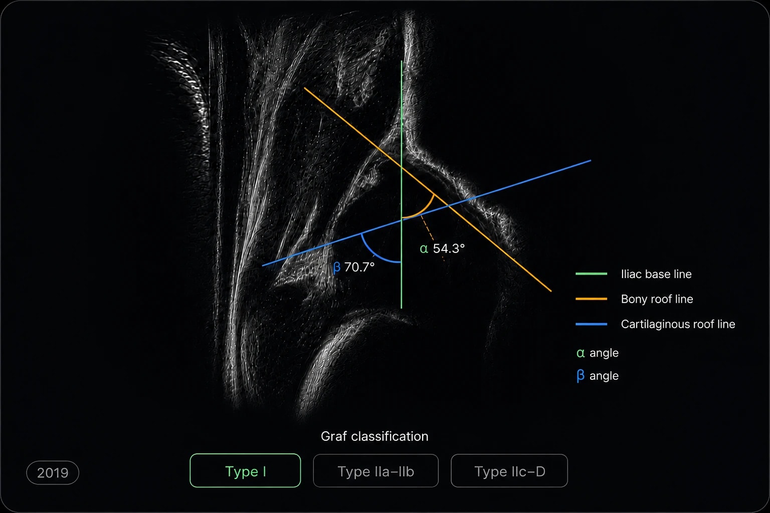
Early foundational study combining automated anatomical structure identification, α–β angle measurement, and classification in a unified computer-aided workflow according to the Graf method.
2020·Ultrasound in Medicine & Biology
Aysun Sezer · Hasan Basri Sezer
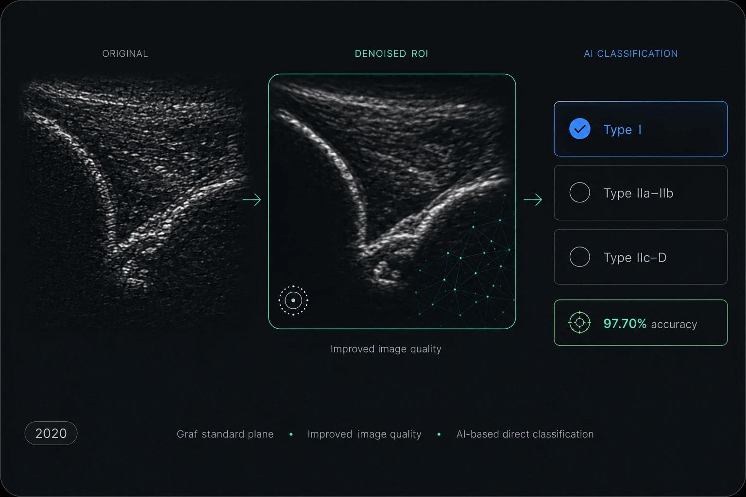
Presents automated AI classification of newborn hip ultrasound images in the Graf standard plane and a novel data augmentation approach based on speckle noise reduction.
2023·Joint Diseases and Related Surgery
Aysun Sezer · Hasan Basri Sezer
A validated segmentation study focused on determining the measurable Graf standard plane through simultaneous automated evaluation of the iliac wing, labrum, and acetabular region.